
Central wrist pain
A deep ache may worsen with activity.
Kienböck’s disease occurs when the lunate bone loses its blood supply and gradually weakens or collapses. Early diagnosis and accurate staging help determine whether the wrist can be unloaded, the lunate can be preserved or a salvage procedure is needed.

Loss of blood supply to the lunate, one of the eight carpal bones.
X-rays may become abnormal later; MRI can detect earlier disease.
Reduce load, preserve the lunate where possible and maintain useful function.
Choice depends heavily on stage, anatomy and surrounding cartilage.
The lunate is a central carpal bone that helps transfer load through the wrist. In Kienböck’s disease its blood supply is disrupted, leading to osteonecrosis, weakening and possible collapse.
The cause is not always clear. Anatomy, repetitive load and previous injury may contribute. Treatment is stage-specific and should be planned by a wrist specialist.
Symptoms vary with the injured wrist structure and how long function has been affected. Persistent pain, clicking, stiffness, weakness or instability deserves specialist assessment.
A deep ache may worsen with activity.
The back of the wrist can appear full.
Flexion and extension may become restricted.
Pain and carpal change reduce strength.
The centre-back of the wrist may be sensitive.
Pain can become more constant as collapse advances.
A useful consultation connects pain location, stability, imaging and function to the exact wrist structure—not just the label.
Forearm-bone length and lunate shape can alter load.
Repeated loading may contribute in susceptible wrists.
Trauma may disrupt blood flow in some patients.
Many patients have no single identifiable cause.
Diagnosis requires staging: whether the lunate is only abnormal on MRI, has become sclerotic, has collapsed, or has led to secondary arthritis.
Pain, motion, grip and tenderness are recorded.
Density, fracture, collapse and carpal alignment are assessed.
MRI can detect early blood-supply change and cartilage condition.
CT can define fracture and collapse for surgical planning.
Surgery is not automatic. Treatment is matched to pain, stability, cartilage condition, imaging and the patient’s goals.

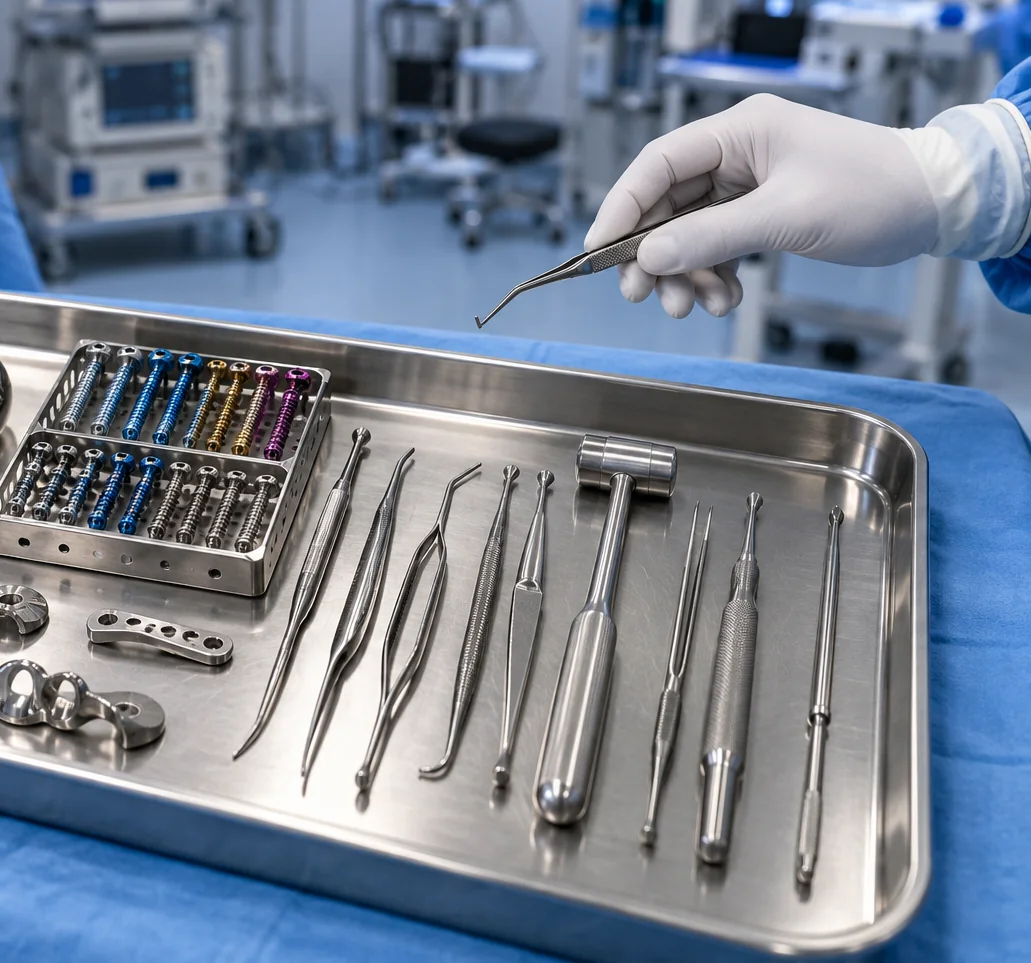
There is no single operation for every stage. The plan depends on bone anatomy, lunate collapse, wrist alignment, cartilage condition, age and activity.
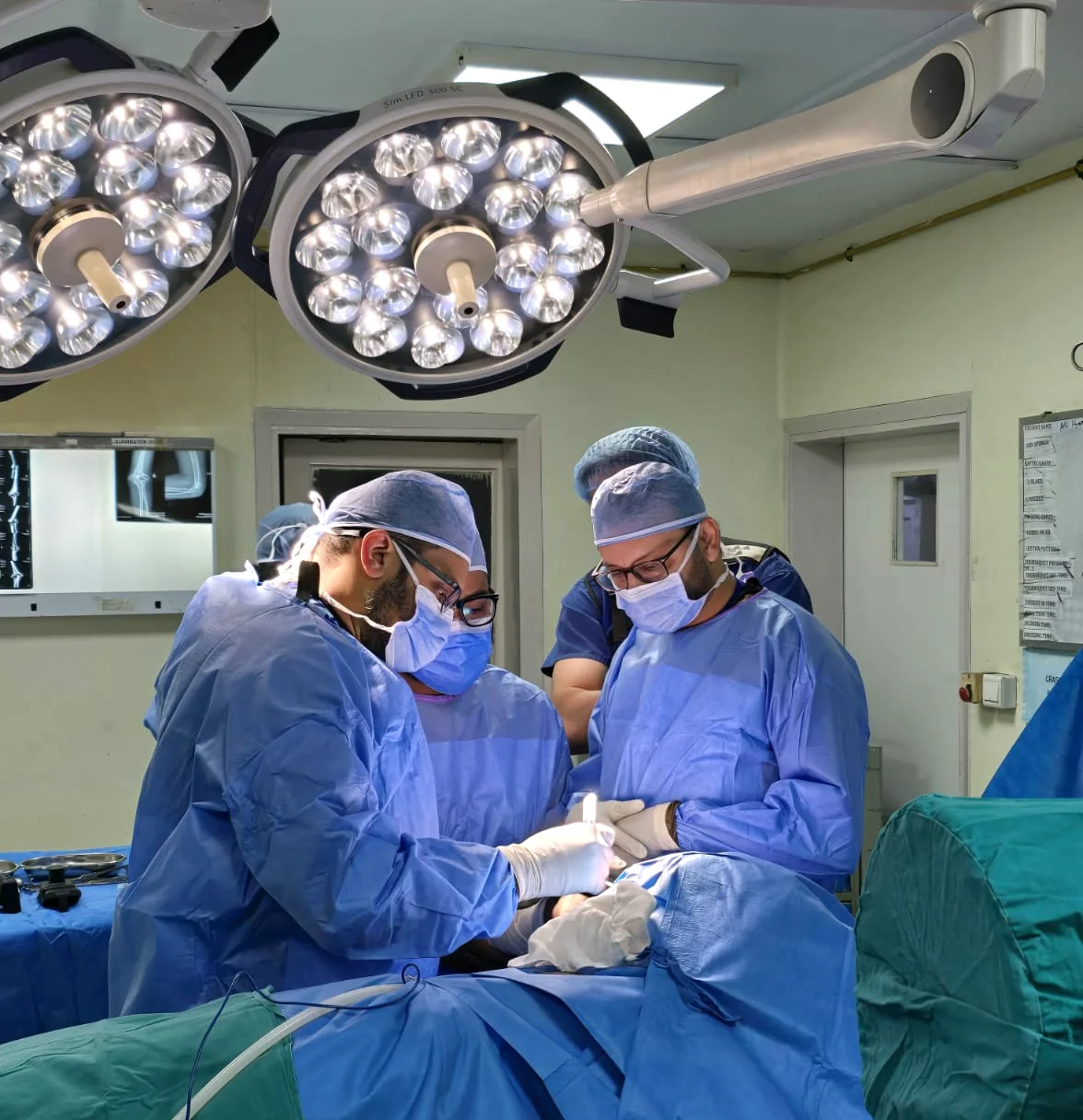
Timelines are guides only. The diagnosis, procedure, stability, cartilage condition, job and associated injuries all influence recovery.
Immobilisation and swelling control depend on the procedure.
Bone healing and wrist motion are reviewed.
Strength and functional use progress gradually.
Recovery and disease monitoring can continue for a year or more.

 4.96/555 verified reviews
Dr Shoaib Arshad is a fellowship-trained consultant orthopaedic hand and wrist surgeon with UK specialist training and more than 17 years in orthopaedic surgery.
General information only. Your diagnosis and treatment plan should be based on an individual medical assessment.
It begins as loss of blood supply to the lunate. Arthritis can develop later if the bone collapses and wrist mechanics change.
Early disease may have a normal X-ray, while MRI can detect changes sooner.
Therapy can help maintain motion and function but does not restore the lunate blood supply.
Each stage has different anatomy and cartilage damage, so treatment ranges from unloading to reconstruction or salvage.
Yes. Dr Arshad can review symptoms and imaging in Manchester, UK and during UK clinical availability.
Request a specialist consultation and bring any previous X-rays, MRI scans, reports, braces or treatment information.