
Visible or palpable lump
A mass may be soft, firm, mobile or fixed.
Most hand and wrist lumps are benign, but the diagnosis should not be guessed from appearance alone. A specialist assessment considers where the lump arises, how it feels, whether it changes and whether imaging or tissue diagnosis is needed.
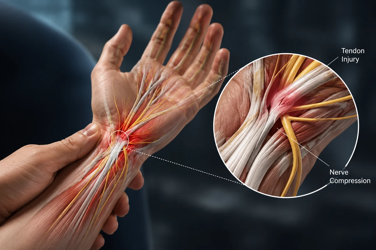
No. Ganglion cysts and benign solid tumours are much more common.
Growth, firmness, pain, skin change, nerve symptoms and whether the mass is fixed.
Clinical examination plus X-ray, ultrasound or MRI when appropriate.
For symptoms, growth, uncertainty or concern about the diagnosis.
Common causes include ganglion cysts, giant cell tumour of tendon sheath, lipoma, epidermal inclusion cyst, carpal boss and benign bone lesions. Malignant tumours are uncommon but must be considered when features are atypical.
The safest approach is to identify whether a mass is fluid-filled, solid, arising from bone, tendon sheath, joint, nerve or skin before deciding on observation or removal.
Symptoms vary with the condition, joint and duration. Persistent pain, stiffness, deformity, a growing lump or loss of function deserves specialist assessment.
A mass may be soft, firm, mobile or fixed.
Ganglions can fluctuate; solid tumours often grow more steadily.
A mass may irritate a tendon or joint.
Pressure on a nearby nerve can alter sensation.
Surface change may provide diagnostic clues.
A mass can mechanically block tendon or joint motion.
A useful consultation connects the symptom pattern to the joint or tissue involved and the underlying cause—not just the label.
Fluid-filled swelling linked to a joint or tendon sheath.
A usually benign solid mass arising around a tendon sheath.
Benign fatty or skin-derived lumps.
Enchondroma and rarer lesions may arise from bone or soft tissue.
The goal is to establish the tissue of origin and identify any warning features before treatment.
Duration, change, pain, trauma and systemic symptoms are reviewed.
Size, consistency, mobility, skin, nerve and tendon relationships are assessed.
These can distinguish bone involvement and fluid-filled from solid masses.
Atypical, deep or concerning lesions may require advanced imaging and tissue diagnosis.
Surgery is not automatic. Treatment is matched to pain, joint or tissue damage, function, medical health and the patient’s goals.
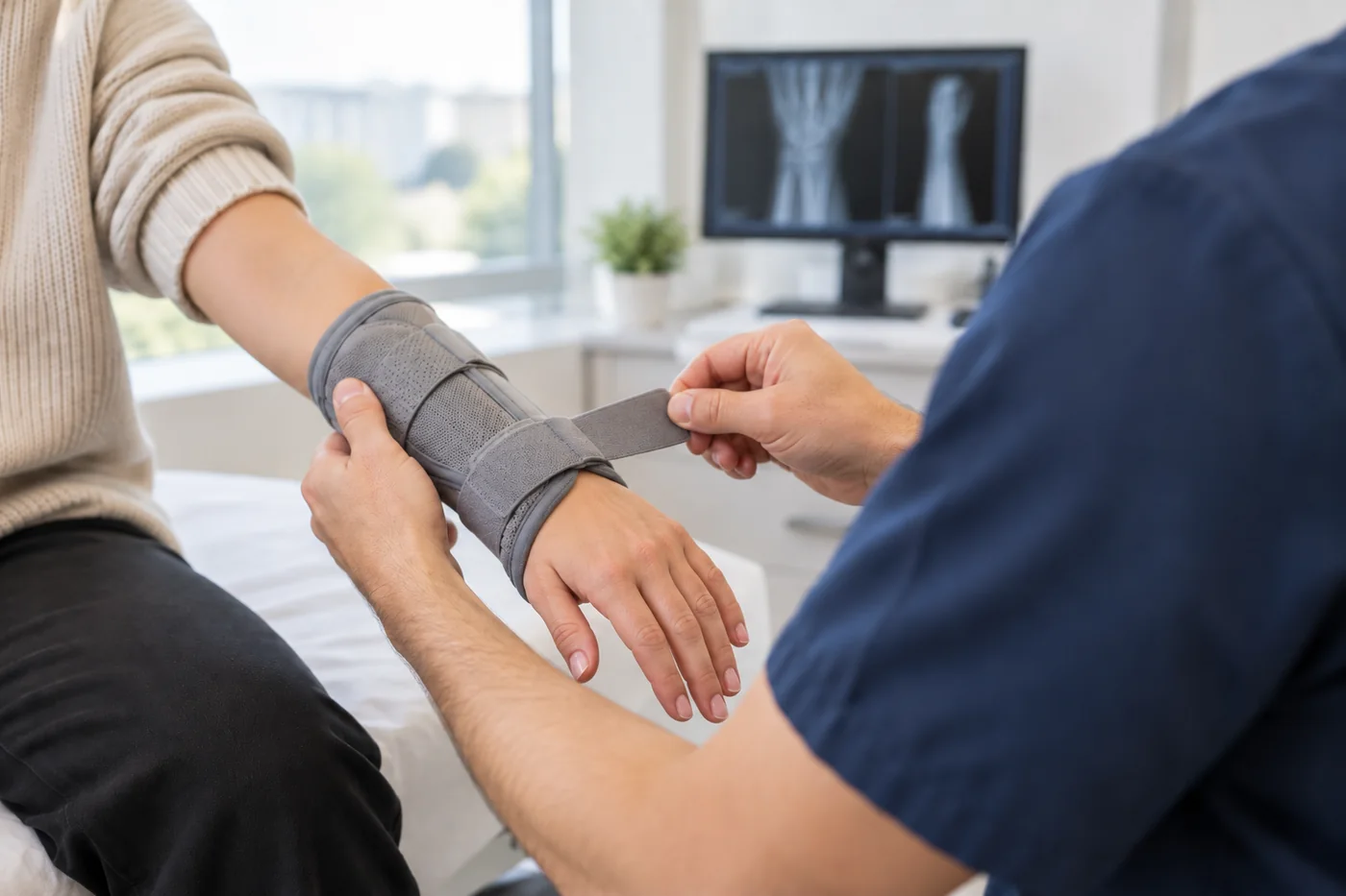
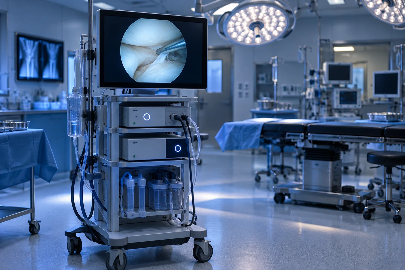
Surgery is considered when a lump is painful, growing, function-limiting, recurrent or diagnostically uncertain. The operation should be planned around the tissue of origin and nearby structures.
Timelines are guides only. The diagnosis, procedure, joint or tissue condition, job and medical health all influence recovery.
Elevation, dressing care and movement of uninvolved joints.
Wound review and pathology discussion when tissue was sent.
Scar care, motion and strength progress according to the site.
Monitor for recurrence or any new symptoms.

 4.96/555 verified reviews
Dr Shoaib Arshad is a fellowship-trained consultant orthopaedic hand and wrist surgeon with UK specialist training and more than 17 years in orthopaedic surgery.
General information only. Your diagnosis and treatment plan should be based on an individual medical assessment.
Most hand and wrist masses are benign, but examination and imaging are needed when the diagnosis is uncertain.
Ultrasound can often distinguish cystic from solid lesions and show their relationship to tendons, but MRI or biopsy may still be needed.
No. A typical painless benign lesion can often be observed.
Removed solid or uncertain tissue is commonly sent for histopathology to confirm the diagnosis.
The team can arrange specialist assessment in Manchester, UK and advise on Dr Arshad’s UK availability.
Request a specialist consultation and bring any previous X-rays, scans, reports or treatment information.