
Unable to bend a finger joint
Loss of active bending is a key warning sign.
A deep cut on the palm side of the hand or finger can damage the tendons that bend the digits. Inability to bend a finger, altered sensation or an open wound needs prompt specialist assessment because repair timing and protected rehabilitation can affect recovery.
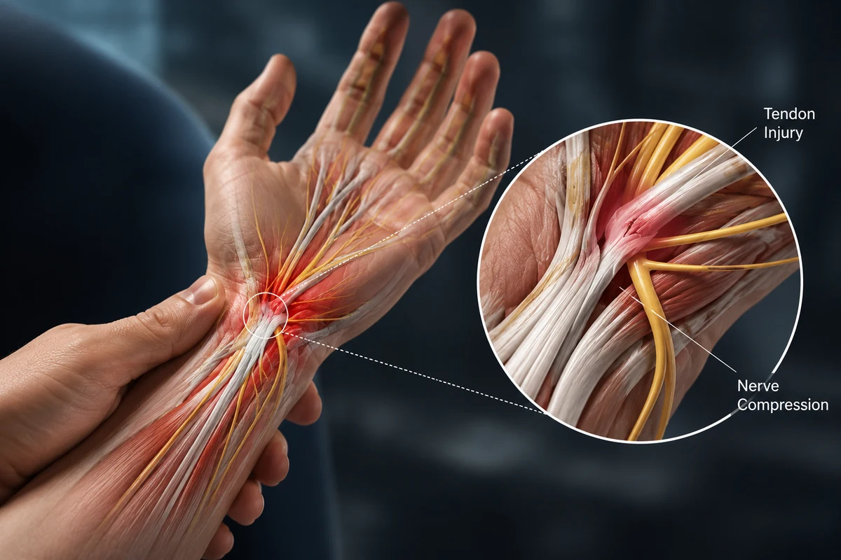
One or more flexor tendons that bend the finger or thumb.
Open cuts, inability to bend, altered feeling, heavy contamination or circulation concerns.
Partial injuries may be managed selectively; complete lacerations usually require repair.
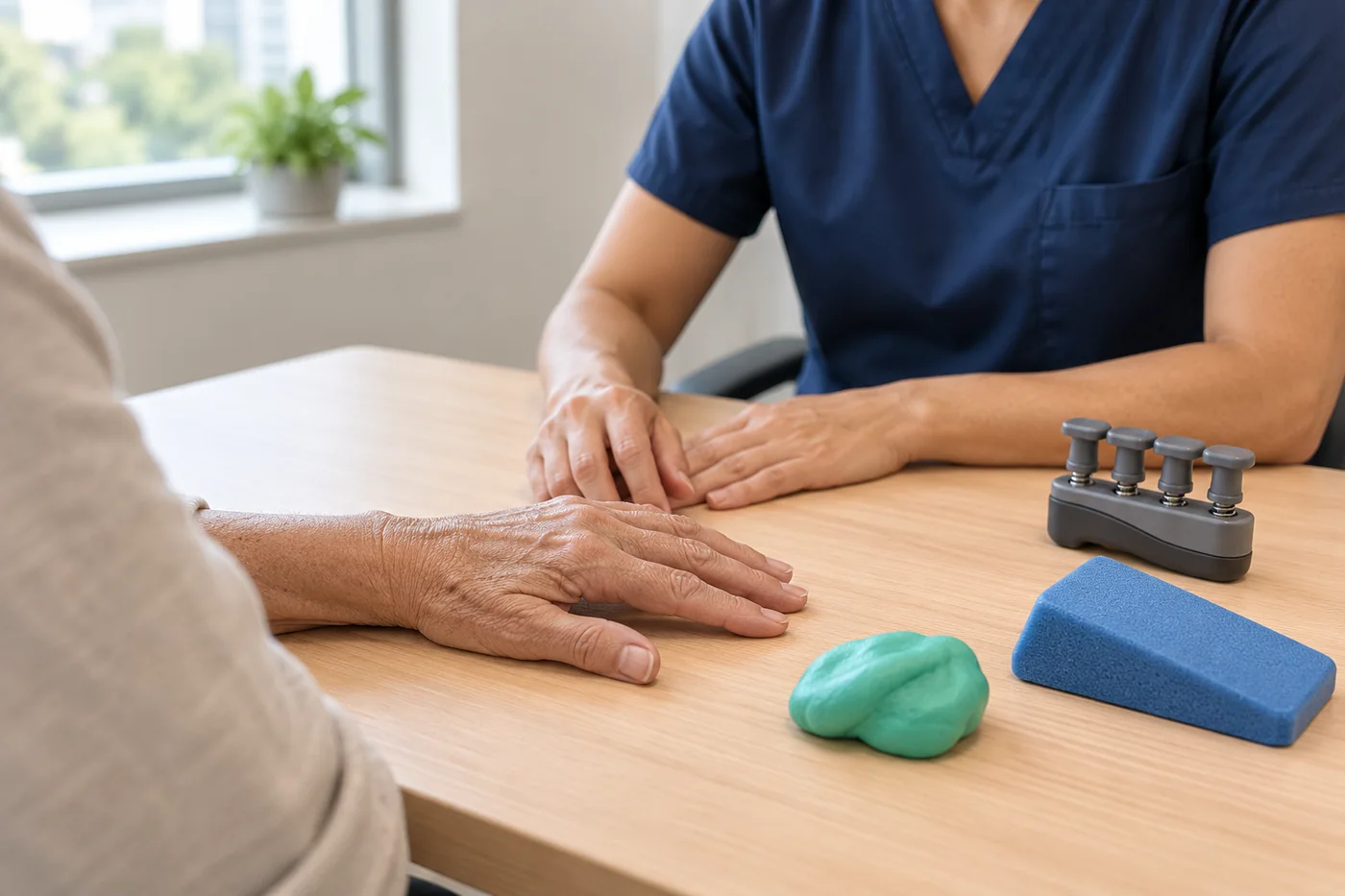
A splint and supervised hand therapy protect the repair while movement is gradually restored.
Flexor tendons run along the palm side of the hand and fingers and connect forearm muscles to the finger bones. They allow the fingers and thumb to bend.
Sharp cuts can partly or completely divide a tendon. Crush injuries, avulsions and fractures may also damage tendons, nerves and blood vessels at the same time.
Symptoms vary with the tendon or finger structure involved and how long function has been affected. Persistent locking, weakness, deformity or loss of movement deserves specialist assessment.
Loss of active bending is a key warning sign.
A partial tendon injury may cause painful or weak flexion.
Cuts near the finger or palm can injure tendon, nerve and vessel together.
The injured finger may sit differently from the others.
Associated digital nerve injury may alter feeling on one side of the finger.
Partial tendon damage can cause swelling, catching or reduced glide.
A useful consultation connects the symptom pattern to the exact tendon, pulley or joint involved—not just the label.
Glass, knives, metal and workplace tools can divide a tendon.
A fingertip may be forcefully straightened while actively gripping, pulling the tendon from the bone.
High-energy injury can damage tendon, bone, nerve and blood vessels.
Less commonly, tendon wear or inflammatory disease can lead to rupture.
Assessment documents each finger joint, tendon function, sensation, circulation, wound contamination and associated bone or joint injury.
Mechanism, timing, position and depth of the wound are important.
Each flexor tendon is tested separately while other joints are stabilised.
Sensation and blood flow are checked before local anaesthetic when possible.
X-ray looks for fracture or foreign body; ultrasound may help in selected cases.
Surgery is not automatic. Treatment is matched to the exact tendon or finger problem, symptom severity, function and the patient’s goals.
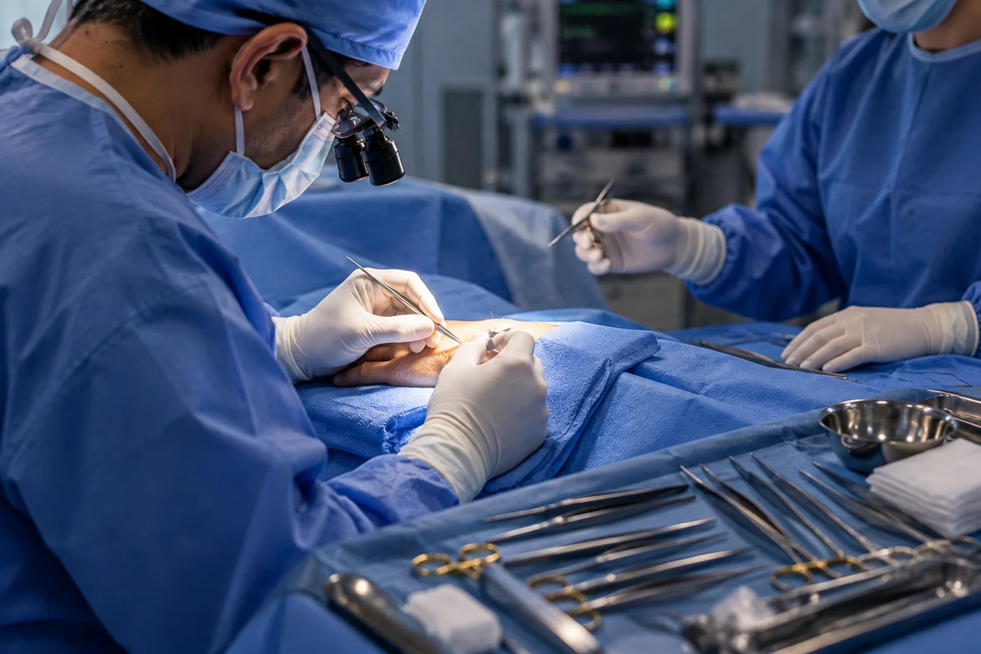
Complete flexor tendon lacerations generally need surgical repair. Timing, wound condition and associated injuries determine the exact plan.
Timelines are guides only. The diagnosis, procedure, wound, therapy programme, job and associated injuries all influence recovery.
The hand is protected in a splint and therapy begins according to the repair protocol.
Movement is carefully controlled; the splint is worn as directed.
Light function and strengthening progress gradually, depending on the repair and therapist advice.
Strength and glide continue to improve; heavy use and sport require individual clearance.

 4.96/555 verified reviews
Dr Shoaib Arshad is a fellowship-trained consultant orthopaedic hand and wrist surgeon with UK specialist training and more than 17 years in orthopaedic surgery.
General information only. Your diagnosis and treatment plan should be based on an individual medical assessment.
Complete lacerations usually require repair. Selected partial injuries may be treated without surgery if function and tendon stability are satisfactory.
Therapy protects the repair while introducing enough controlled movement to reduce stiffness and tendon adhesions.
Repairs remain weak for weeks and commonly take around three months to regain substantial strength. Timelines vary by zone and injury.
Yes. Digital nerves run close to the flexor tendons and can be injured by the same cut.
The team can arrange Manchester, UK consultation and advise on Dr Arshad’s UK availability.
Request a specialist consultation and bring any previous imaging, reports, splints or treatment information.