
Loss of active extension
The finger, thumb or wrist cannot be fully straightened.
A cut or impact to the back of the hand can damage the tendons that straighten the fingers. Drooping, loss of extension or an open wound should be assessed promptly so that splinting, repair and therapy can be planned before stiffness or deformity develops.
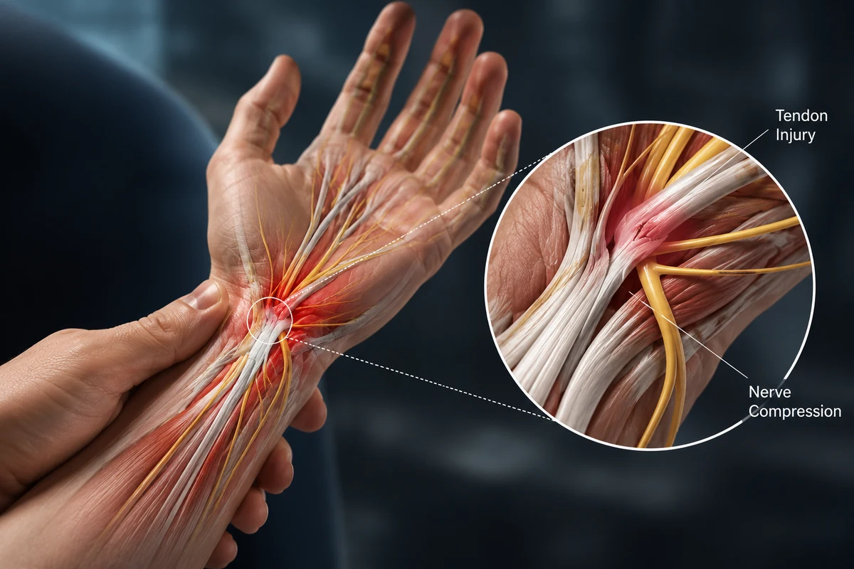
The thin extensor tendons that straighten the finger, thumb or wrist.
A finger or joint cannot be actively straightened even though it may be moved passively.
Splinting for selected closed injuries or repair for open/complete lacerations.
Protected movement and hand therapy continue for weeks while the repair regains strength.
Extensor tendons lie close beneath the skin on the back of the hand and fingers. They straighten the joints and coordinate complex finger movement through the extensor mechanism.
Because they are superficial, they can be injured by relatively small cuts. Closed impacts can also tear or detach tendon structures, leading to mallet finger or boutonnière deformity.
Symptoms vary with the tendon or finger structure involved and how long function has been affected. Persistent locking, weakness, deformity or loss of movement deserves specialist assessment.
The finger, thumb or wrist cannot be fully straightened.
The fingertip or middle joint may rest in a bent position.
Even a small cut can divide the superficial tendon.
Tenderness may follow a direct blow or laceration.
The joint can be moved straight passively but cannot hold the position actively.
Untreated central slip injury may progress to boutonnière deformity.
A useful consultation connects the symptom pattern to the exact tendon, pulley or joint involved—not just the label.
Glass, metal or a knife can divide the tendon on the back of the hand.
A ball or impact can force a finger joint to bend and rupture the tendon.
Bone injury may detach the tendon or disrupt the extensor mechanism.
Arthritis or chronic tendon wear can cause delayed rupture.
The examination identifies which extensor zone is involved, tests each joint separately and checks for fracture, joint instability, nerve injury and wound contamination.
The position of the cut or impact predicts which tendon structure may be injured.
Each joint is assessed for extension strength and lag.
Targeted manoeuvres can help assess the central slip and lateral bands.
Imaging may identify fracture, joint alignment, foreign body or tendon discontinuity.
Surgery is not automatic. Treatment is matched to the exact tendon or finger problem, symptom severity, function and the patient’s goals.
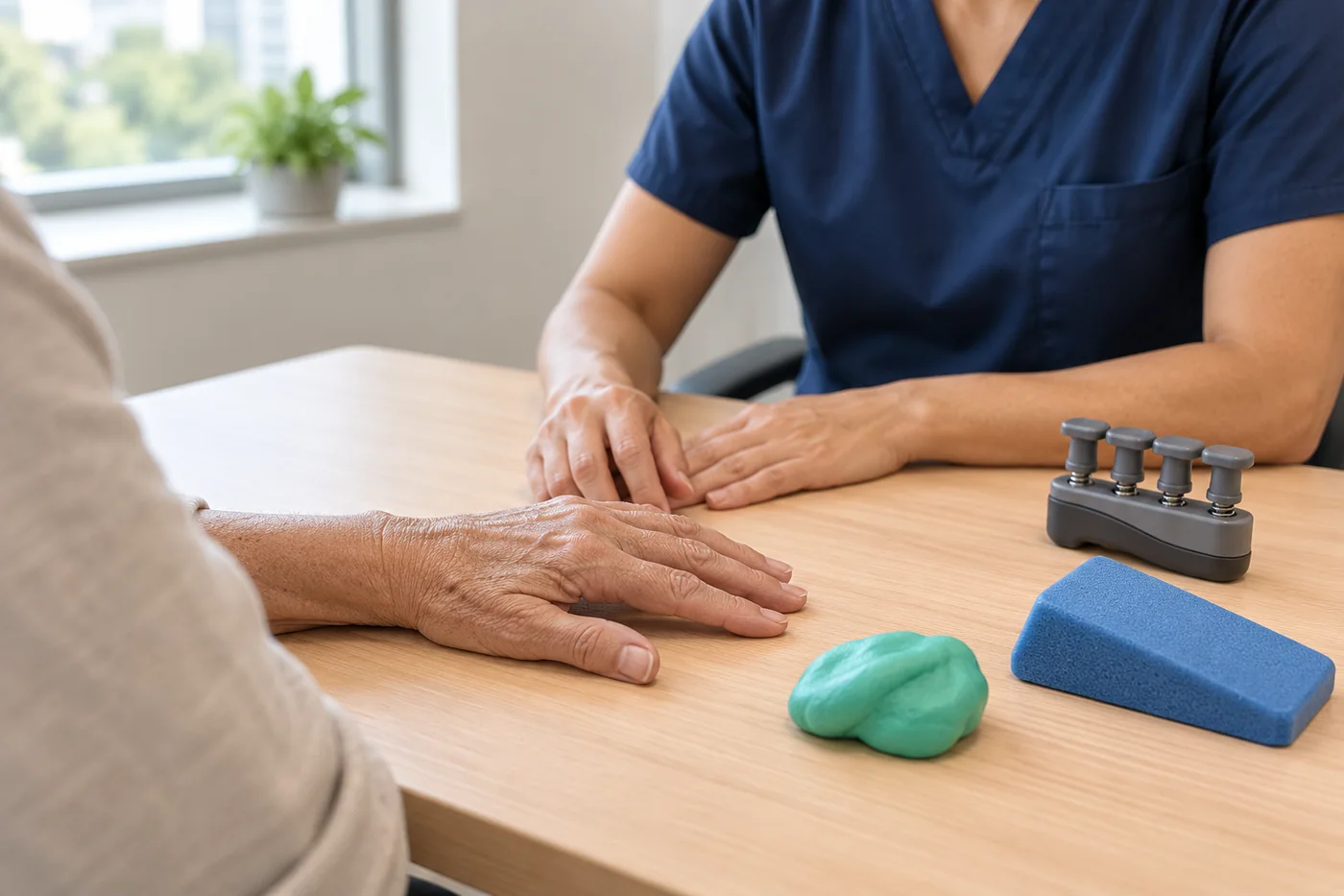

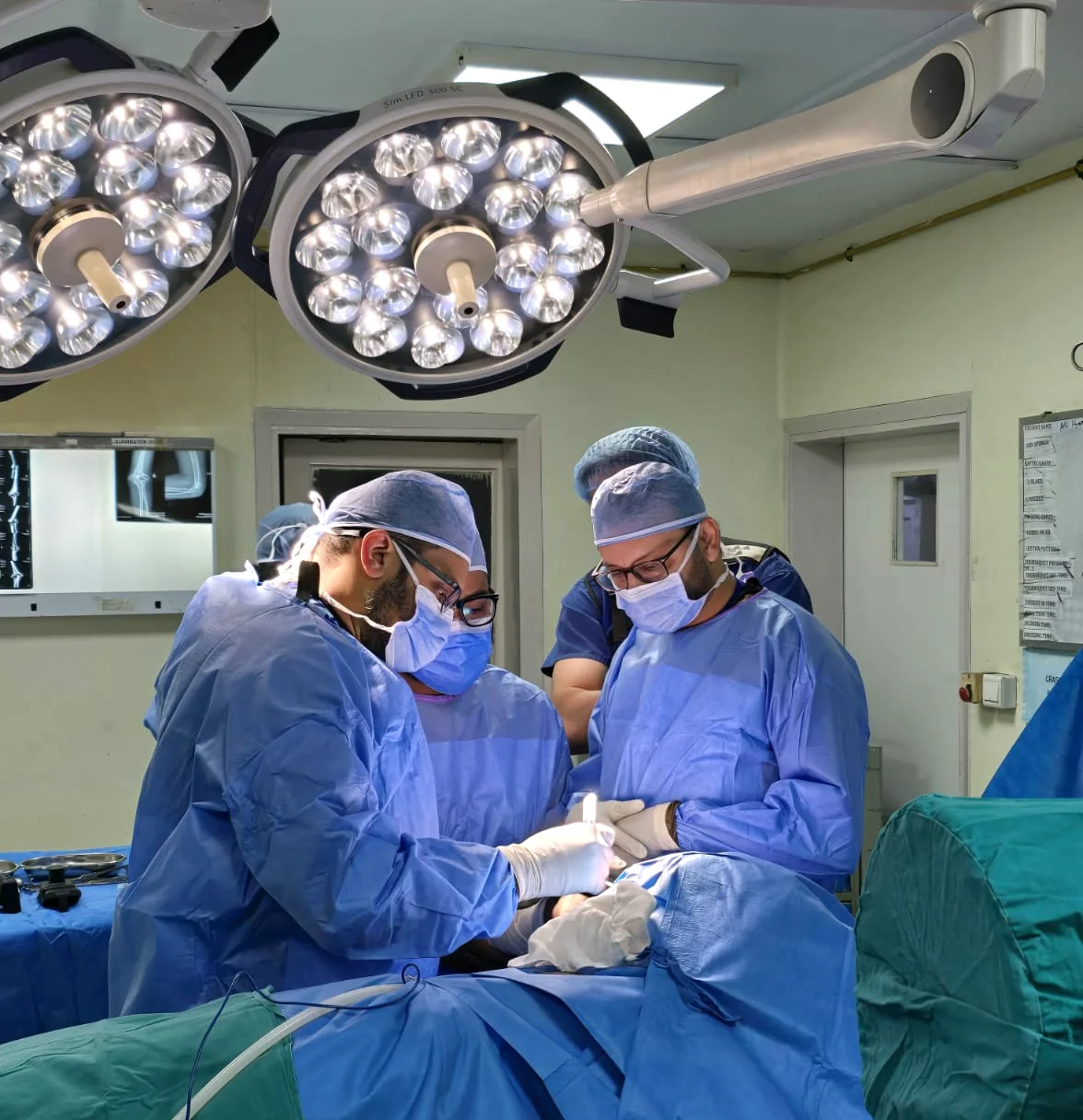
Surgery is commonly required for complete open lacerations and selected displaced fractures, unstable injuries or failed splint treatment.
Timelines are guides only. The diagnosis, procedure, wound, therapy programme, job and associated injuries all influence recovery.
The repair is protected in a splint and the wound is monitored.
Exercises are carefully controlled by the hand therapist and depend on the repair zone.
Movement and light function progress while the tendon continues to regain strength.
Heavier use is introduced only after adequate healing and clinical review.

 4.96/555 verified reviews
Dr Shoaib Arshad is a fellowship-trained consultant orthopaedic hand and wrist surgeon with UK specialist training and more than 17 years in orthopaedic surgery.
General information only. Your diagnosis and treatment plan should be based on an individual medical assessment.
Some closed injuries can heal with precise continuous splinting. Complete open lacerations often need surgical repair.
The tendon heals at a specific length. Allowing the joint to bend can separate the healing ends and lead to a persistent droop.
The tendon commonly remains vulnerable for about three months, though the exact rehabilitation plan depends on the injury zone.
The joint can be straightened passively but cannot hold full extension actively because the tendon mechanism is weak or elongated.
Dr Arshad sees private patients at The Highfield Hospital, Manchester Road, Rochdale OL11 4LZ — within easy reach of Manchester, Oldham, Bury, Bolton and the wider North West. You can book online through Circle Health Group, call +44 7443 213645, or send an enquiry and the team will help.
Request a specialist consultation and bring any previous imaging, reports, splints or treatment information.